

Varicose veins: Diagnosis and treatment from a single source
Introduction
Ultrasound examinations use harmless sound waves to make the inside of the body visible. As radiologists, we use the method frequently and in a variety of ways, for example to examine organs such as the liver, pancreas, kidneys, blood vessels or joints.
The term ultrasound actually comes from physics. It refers to sound waves whose frequency is above the human hearing range and which the human ear can no longer perceive. In medicine, an imaging procedure has been developed from this, which is often colloquially called "ultrasound". Other terms for the examination are sonography or echography.
Ultrasound examinations pose little risk when used correctly.

Herniated disc
When a disc herniates, the tissue of the disc between the vertebral bodies is pushed outwards more and more. Symptoms arise when the bulging disc or protruding tissue presses on a nerve root or nerve. The more the nerve root is compressed, the more severe the symptoms are.
Often, disc damage is caused by years of wear and tear or poor posture without any pain. Then a trigger is enough to bring about the symptoms: a clumsy movement, a sudden twisting of the spine, drafts, dampness, hypothermia, etc. Sudden shooting pain is typical, colloquially known as "lumbago". If nerve structures are trapped by large parts of the disc, sensory disturbances and loss of strength, even paralysis, can also occur.
Depending on the degree of damage to the intervertebral disc, the following forms are distinguished:
Disc protrusion: The disc bulges between the vertebral bodies. The disc's shell is intact.
Herniated disc (extrusion): The outer shell of the disc is torn and the inner core bulges outwards.
Sequestered disc herniation (sequestrum): Tissue from the disc has protruded through the torn sheath into the spinal canal.

Diagnostics
Magnetic resonance imaging (MRI) is the gold standard for diagnosing herniated discs and spinal stenosis. It allows for high-resolution visualization of anatomical structures and enables a reliable diagnosis.
MRI is also used for millimeter-precise planning of the CT-guided infiltrations we perform. Our patients usually bring their MRI scans to our office on a data carrier, and we then explain the images and plan the procedure. Alternatively, we can arrange an MRI scan for our patients.
An MRI scan of the lumbar or cervical spine takes approximately 15 minutes and does not involve harmful X-rays. As board-certified radiologists, we are qualified by our training and professional regulations to interpret and report these examinations.
Therapy
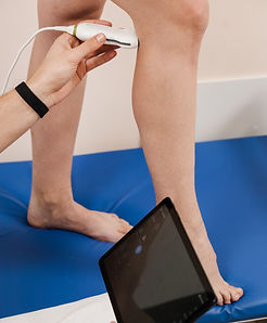
As a private practice for phlebology in Vienna, we specialize in minimally invasive catheter treatments for varicose veins and spider veins. These methods leave no scars or incisions and allow for a gentle and almost painless treatment.
First, the affected vein is visualized using ultrasound. Then a laser probe catheter is inserted into the great saphenous vein through a small skin incision.
As the probe is withdrawn, we activate the laser, which damages the vein wall and causes it to stick together. This closes the vein and the blood flow is taken over by surrounding healthy veins
We locate the veins precisely using a special light source and apply a sclerosing agent using a special needle. This irritates and sticks together the inner wall of the vein (intima). The treated vessels stick together from the inside, close and are broken down by the body within a few weeks. This results in an attractive cosmetic result.
Results before and after therapy

