
CT-guided injection:
Diagnostics and therapy
Introduction
Ultrasound examinations use harmless sound waves to make the inside of the body visible. As radiologists, we use the method frequently and in a variety of ways, for example to examine organs such as the liver, pancreas, kidneys, blood vessels or joints.
The term ultrasound actually comes from physics. It refers to sound waves whose frequency is above the human hearing range and which the human ear can no longer perceive. In medicine, an imaging procedure has been developed from this, which is often colloquially called "ultrasound". Other terms for the examination are sonography or echography.
Ultrasound examinations pose little risk when used correctly.

Herniated disc
When a disc herniates, the tissue of the disc between the vertebral bodies is pushed outwards more and more. Symptoms arise when the bulging disc or protruding tissue presses on a nerve root or nerve. The more the nerve root is compressed, the more severe the symptoms are.
Often, disc damage is caused by years of wear and tear or poor posture without any pain. Then a trigger is enough to bring about the symptoms: a clumsy movement, a sudden twisting of the spine, drafts, dampness, hypothermia, etc. Sudden shooting pain is typical, colloquially known as "lumbago". If nerve structures are trapped by large parts of the disc, sensory disturbances and loss of strength, even paralysis, can also occur.
Depending on the degree of damage to the intervertebral disc, the following forms are distinguished:
Disc protrusion: The disc bulges between the vertebral bodies. The disc's shell is intact.
Herniated disc (extrusion): The outer shell of the disc is torn and the inner core bulges outwards.
Sequestered disc herniation (sequestrum): Tissue from the disc has protruded through the torn sheath into the spinal canal.

Diagnostics
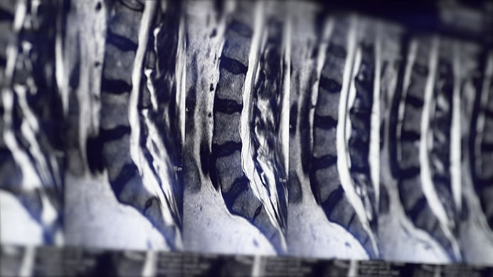
Magnetic resonance imaging (MRI) is the gold standard for diagnosing herniated discs and spinal stenosis. It allows for high-resolution visualization of anatomical structures and enables a reliable diagnosis.
MRI is also used for millimeter-precise planning of the CT-guided infiltrations we perform. Our patients usually bring their MRI scans to our office on a data carrier, and we then explain the images and plan the procedure. Alternatively, we can arrange an MRI scan for our patients.
An MRI scan of the lumbar or cervical spine takes approximately 15 minutes and does not involve harmful X-rays. As board-certified radiologists, we are qualified by our training and professional regulations to interpret and report these examinations.

Therapy
We and our patients have had the best experiences with CT (computed tomography) targeted infiltration in the gentle and effective treatment of herniated discs, spinal stenosis and facet joint arthrosis.
The advantage of this minimally invasive intervention is that the medication is administered precisely to the area causing the pain by precisely positioning the infiltration needle.
For this purpose, we have developed a tailor-made and tried-and-tested drug mixture of glucocorticoids (anti-inflammatory and decongestant effects) and local anesthetics (pain-relieving effects) with long-term effects.
We can thus achieve rapid pain relief and reduction of swelling in cases of herniated discs, spinal canal stenosis (narrowing of the spinal canal) and facet joint arthrosis.